
Coronary Plaque and Atherosclerosis: What Treatment Can Really Achieve
Plaque builds inside the artery wall over decades, and the artery enlarges outward to accommodate it — which is why significant disease can be present while stress tests and angiograms look normal. Treatment does produce real regression, but modestly: the best trials shrink plaque by roughly 1–2%. What changes dramatically is the plaque’s composition — from soft and rupture-prone to dense, calcified and stable — and that is what saves lives. Your arteries are not a clogged pipe that can be cleaned out. They are living tissue that scars and heals.
What Is Coronary Artery Plaque?
Your coronary arteries are the blood vessels that wrap around your heart and deliver oxygen-rich blood to the heart muscle. They are about the width of a drinking straw — roughly 3 to 4 millimeters across. Coronary artery plaque is a buildup of cholesterol, fat, inflammatory cells, calcium, and scar tissue inside the walls of these arteries. Over decades, this buildup can narrow the artery, reduce blood flow to the heart, or — in the worst case — rupture suddenly and cause a heart attack.
Understanding how plaque forms, grows, and changes is one of the most important things you can do for your heart health. This guide walks you through the entire life cycle of a plaque — from the very first invisible changes in childhood to the advanced disease that causes heart attacks — and explains what modern medicine can and cannot do to reverse it. If you want to know how your arteries are ageing overall, start with your vascular age.
Part 1: How Plaque Forms and Grows
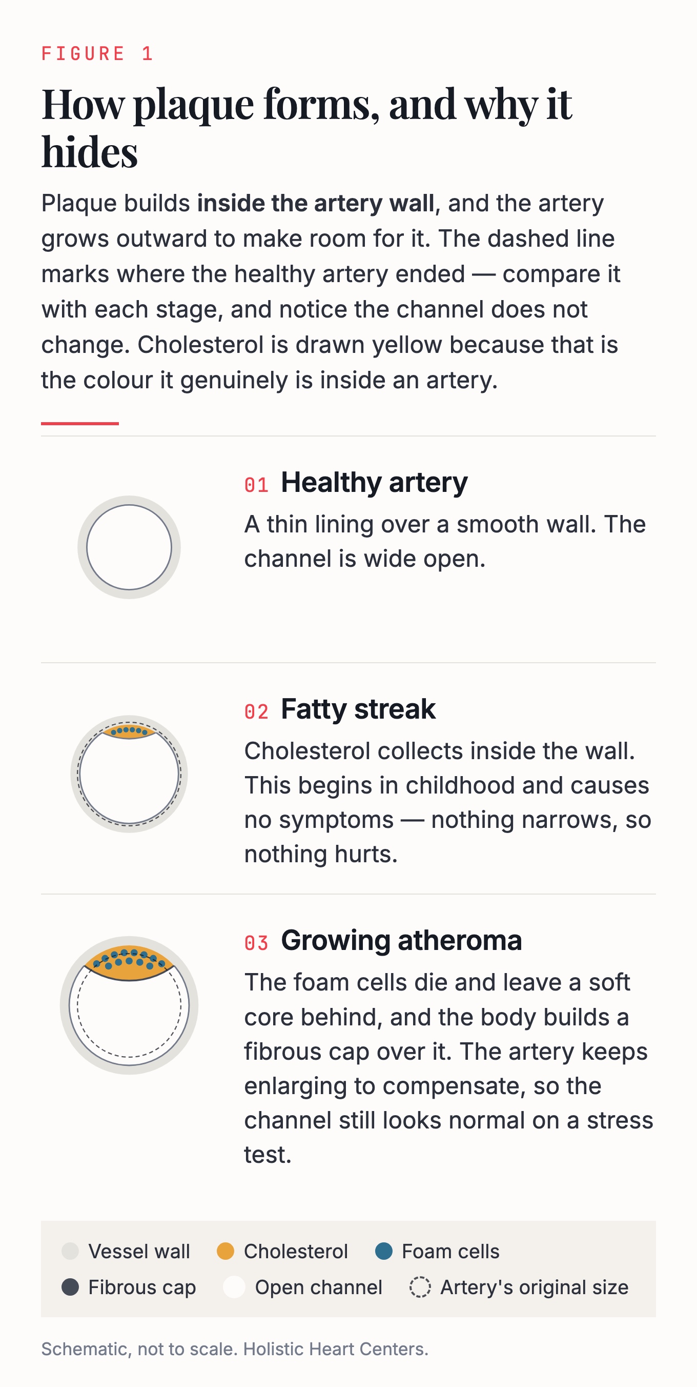
Figure 1. Plaque builds inside the artery wall, and the artery grows outward to make room for it. The channel stays open long after the disease has started.
Stage 1: The Invisible Beginning (Childhood Through Young Adulthood)
Plaque formation begins far earlier than most people realize. Autopsy studies of children and young adults have shown that fatty streaks — the earliest form of plaque — are present in the arteries of nearly all children by their teenage years. By the time a person reaches their 20s and 30s, more advanced deposits can already be forming, especially in those who smoke, have high cholesterol, or are overweight.
At this stage, there are absolutely no symptoms. No test your doctor can order will detect these changes (except in research settings). The artery looks and functions normally from the outside. But inside the artery wall, cholesterol particles — particularly LDL cholesterol, often called “bad cholesterol” — are seeping into the inner lining of the vessel and triggering an inflammatory response.
Think of it like water damage behind a wall in your house. The wall looks fine from the outside, but behind the paint, moisture is slowly causing damage.
What is happening inside the artery wall:
- LDL cholesterol particles penetrate the inner lining (called the endothelium) and become trapped in the artery wall
- The body’s immune system recognizes these trapped cholesterol particles as foreign invaders
- White blood cells called monocytes are recruited to the area, where they transform into macrophages — essentially “cleanup crews” that try to eat the cholesterol
- These macrophages become so engorged with cholesterol that they turn into “foam cells” — bloated, dysfunctional cells that cannot escape the artery wall
- The accumulation of foam cells creates a visible fatty streak beneath the surface
Stage 2: Silent Growth (30s Through 50s)
As risk factors accumulate over the years — high blood pressure, high cholesterol, diabetes, smoking, obesity, sedentary lifestyle — the plaque grows larger and more complex. This is still a silent process. Most people at this stage feel completely fine.
During this phase, something remarkable happens: the artery actually expands outward to accommodate the growing plaque, like a balloon inflating. This is called “positive remodeling.” Because the artery gets bigger, the channel for blood flow (the lumen) stays the same size, and blood flow is not restricted. This is why stress tests and even traditional angiograms can look completely normal even when significant plaque is present.
What is happening inside the artery wall:
- The fatty streak evolves into a more complex structure called an atheroma
- A pool of dead cells, cholesterol crystals, and debris forms in the center — this is called the necrotic core (think of it as a pocket of “sludge” inside the plaque)
- The body tries to wall off this dangerous material by building a fibrous cap over it — a layer of tough scar-like tissue made of collagen and smooth muscle cells
- New tiny blood vessels grow into the plaque from the outer wall of the artery (like roots growing into soil), which can leak and cause bleeding inside the plaque
- Calcium begins to deposit in the plaque, similar to how calcium deposits form in aging joints or tendons
At this stage, a coronary artery calcium (CAC) scan — a simple, low-radiation CT scan — may detect the first signs of calcification, providing the earliest clinical evidence that plaque is present. Calcium score and coronary CTA explains how that testing works.
Stage 3: The Stable Plaque (Typical Angina Patient)
If plaque continues to grow, it eventually becomes large enough to narrow the artery and restrict blood flow. When the narrowing exceeds about 50–70%, the heart muscle may not get enough blood during exercise or stress, causing chest pain or pressure called angina.
This is the classic “stable plaque.” It has a thick, strong fibrous cap, significant calcification, and a relatively small or dried-out necrotic core. Paradoxically, this type of plaque is actually relatively safe — the thick cap and dense calcium make it very unlikely to rupture. The main problem is that it restricts blood flow, causing symptoms with exertion. Living with calcified arteries goes into what that means day to day.
What is happening inside the artery wall:
- The fibrous cap is thick (well over 65 micrometers — about the width of a human hair)
- The necrotic core has been largely replaced by dense fibrous tissue and sheets of calcium
- Inflammatory cells are relatively sparse — the “fire” has largely burned out
- The artery can no longer expand outward to compensate, so the plaque now encroaches on the blood flow channel
Stage 4: The Dangerous Plaque (The “Ticking Time Bomb”)
This is the most important stage to understand, because it is the one that causes most heart attacks — and it is the most counterintuitive.
The most dangerous plaques are often NOT the ones that cause the most narrowing. Instead, they are plaques that have a large, soft, lipid-rich core covered by a very thin fibrous cap — less than 65 micrometers thick. These are called “thin-cap fibroatheromas” or “vulnerable plaques.” Because the artery has expanded outward to accommodate them, they may cause little or no narrowing on an angiogram. A stress test may be completely normal.
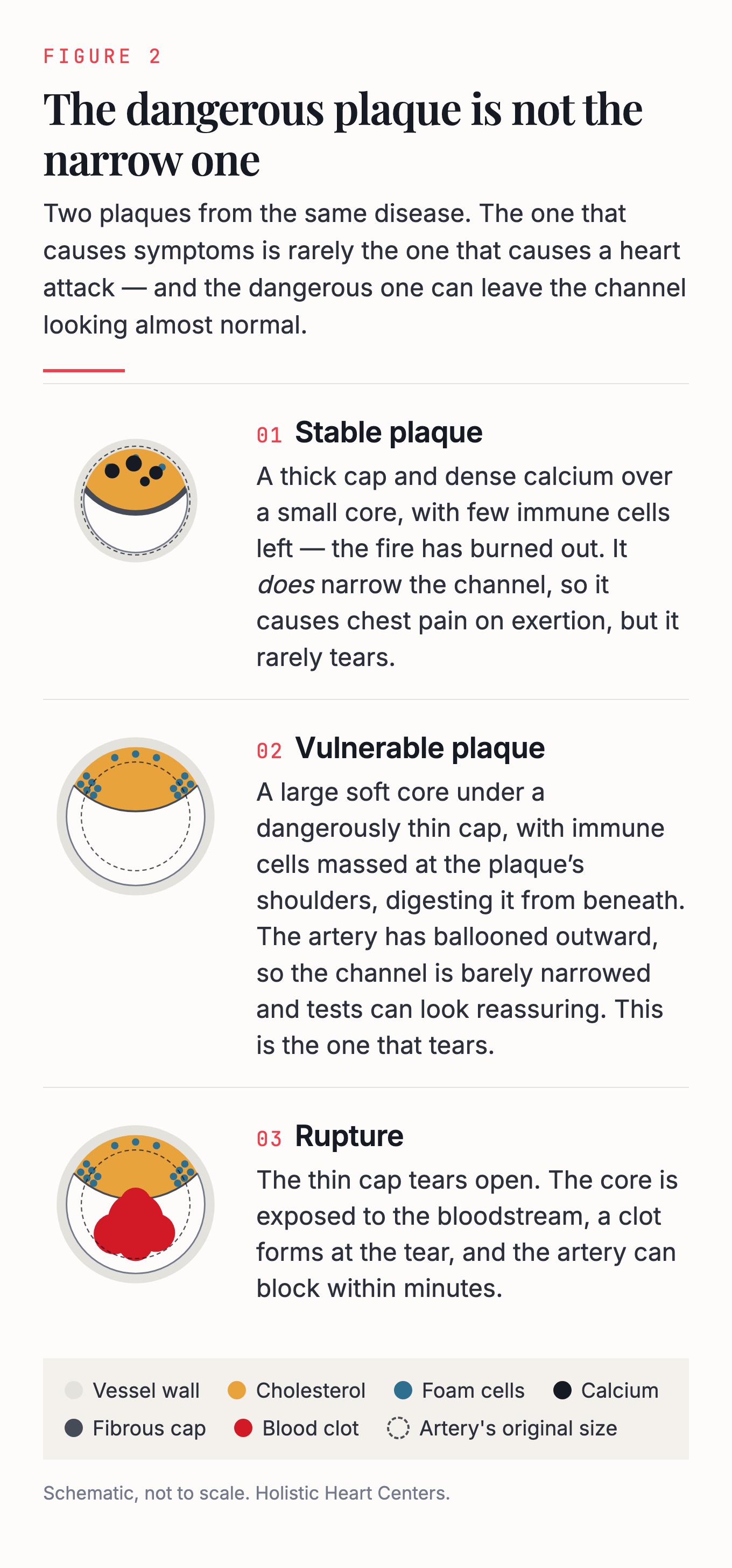
Figure 2. The plaque that causes symptoms is rarely the plaque that causes the heart attack.
Yet these are the plaques that rupture. When the thin cap tears open, the contents of the necrotic core are exposed to the bloodstream, triggering a massive blood clot that can completely block the artery within minutes. This is a heart attack.
What is happening inside the artery wall:
- The necrotic core is large — occupying 30–40% or more of the plaque
- The fibrous cap is dangerously thin — less than 65 micrometers
- Inflammatory cells (macrophages) are concentrated at the “shoulders” of the plaque, actively digesting the cap with enzymes
- Tiny spotty calcifications (microcalcifications) are scattered through the cap, creating stress points — like perforations in a piece of paper that make it easier to tear
- The plaque is “positively remodeled” — the artery has ballooned outward, hiding the plaque from standard tests
People with diabetes and metabolic syndrome are particularly prone to having multiple vulnerable plaques throughout their coronary arteries.
Stage 5: The Heart Attack (Plaque Rupture or Erosion)
When a vulnerable plaque ruptures, the body’s clotting system is activated instantly. A blood clot (thrombus) forms at the rupture site and can completely block the artery. The heart muscle downstream is starved of oxygen and begins to die. This is a heart attack (myocardial infarction). For the bigger picture of the disease itself, see coronary artery disease.
There are actually two ways a plaque can cause a heart attack:
- Plaque rupture (about 60–75% of heart attacks): The thin cap tears open, exposing the necrotic core. This tends to occur in older patients with multiple risk factors and high levels of inflammation. It often causes a complete blockage (STEMI — the most severe type of heart attack).
- Plaque erosion (about 25–40% of heart attacks): The surface lining of the plaque wears away without the cap actually tearing. This tends to occur in younger patients, more often women, with fewer traditional risk factors. It often causes a partial blockage (NSTEMI — a less severe but still serious heart attack).
Part 2: How We Measure Plaque
When you hear about plaque “regression” in medical studies, the researchers are using very specific measurements. Understanding these numbers will help you interpret what “plaque reversal” really means.
Percent Atheroma Volume (PAV)
This is the most commonly used measurement in plaque regression studies. It answers the question: “What percentage of the total artery wall is occupied by plaque?”
Imagine slicing the artery into very thin cross-sections, like slicing a sausage. In each slice, you can see the outer wall of the artery and the inner channel where blood flows. The space between them is the artery wall — and plaque lives in this wall. PAV measures what fraction of the total vessel area is taken up by plaque.
A typical PAV in study patients is around 35–40%. This means that roughly 35–40% of the cross-sectional area of the artery wall is occupied by plaque material.
In the most successful plaque regression trials — using the most powerful medications available — PAV decreased by about 1% to 2%. For example, in the PACMAN-AMI trial, patients who received both a high-intensity statin and a PCSK9 inhibitor (a powerful cholesterol-lowering injection) achieved a PAV reduction of 2.13%, with their LDL cholesterol dropping to an average of 23.6 mg/dL.
To put this in perspective: if a patient starts with a PAV of 42%, the most aggressive treatment available might bring it down to about 40%. The plaque is still there — it is just slightly smaller and, more importantly, much safer in its composition.
Total Atheroma Volume (TAV)
This measures the actual physical volume of plaque in a segment of artery, expressed in cubic millimeters (mm³). Think of it as measuring the total amount of plaque material, rather than the percentage. In major trials, TAV reductions of about 4–6 mm³ have been achieved with intensive therapy.
Why These Numbers Matter
These measurements make clear that plaque regression is real but modest in absolute terms. The artery does not return to a pristine, plaque-free state. What changes most dramatically is not the size of the plaque but its composition — and that compositional change is what saves lives.
Part 3: What Actually Happens During Plaque Regression
The Compositional Transformation
When effective treatment is applied — particularly high-intensity statin therapy, with or without additional cholesterol-lowering medications — the plaque undergoes a profound internal transformation. This is the most important concept in understanding plaque regression:
The plaque does not melt away. It transforms from a dangerous, unstable structure into a safer, more stable one.
Here is what happens inside the plaque during regression:
- The lipid core shrinks. Statins and other cholesterol-lowering drugs reduce the amount of LDL cholesterol circulating in the blood. With less cholesterol being deposited into the plaque, and with the body’s cleanup mechanisms still working, the soft, lipid-rich core gradually gets smaller. In the YELLOW trial, high-dose rosuvastatin reduced the lipid content of plaques by 32% in just 7 weeks.
- The fibrous cap thickens. As inflammation decreases, smooth muscle cells migrate into the cap and produce collagen, making it thicker and stronger. Studies using optical coherence tomography (OCT) — a high-resolution imaging technique — have shown that statin therapy increases fibrous cap thickness by 20 micrometers or more. A thicker cap means the plaque is much less likely to rupture.
- Inflammatory cells decrease. The macrophages that were actively digesting the cap and destabilizing the plaque become less numerous and shift from an aggressive “M1” inflammatory state to a calmer “M2” healing state. This is like switching from a demolition crew to a repair crew.
- Calcification increases. This is the part that confuses many patients. As the plaque heals, the scattered, dangerous microcalcifications coalesce into larger, denser sheets of calcium. This dense calcium actually stabilizes the plaque — like reinforcing a crumbling wall with concrete. This is why your coronary calcium score may go UP even as your plaque becomes safer. A rising calcium score on statin therapy is generally a sign of plaque healing, not worsening disease.
- The artery remodels inward. As the plaque shrinks slightly, the artery wall contracts inward (negative remodeling), and the blood flow channel may modestly enlarge.
The Calcium Paradox
One of the most confusing aspects of plaque treatment for patients is the “calcium paradox.” Your doctor may tell you that your coronary calcium score has increased, even though you have been faithfully taking your statin and your cholesterol is well controlled. This can feel alarming — but it is actually expected and, in most cases, a sign that treatment is working.
Here is why: Statins do not create new plaque. They modify existing plaque. Part of that modification involves converting soft, dangerous, lipid-rich plaque into dense, stable, calcified plaque. The calcium score measures the total amount of calcium in your arteries, so as soft plaque converts to calcified plaque, the score goes up — even though the total amount of plaque has slightly decreased and the remaining plaque is much safer.
If you had a wall made of wet sand (dangerous, could collapse at any time), and you replaced some of that sand with concrete (stable, strong), the wall would actually be safer — even though it now contains a harder material.
What Does NOT Happen During Regression
This is where the popular narrative of “plaque reversal” can be misleading:
- The artery does not return to normal. Even with the most aggressive treatment, the vast majority of plaque volume remains. The artery wall retains a permanent structural footprint of the disease — fibrosis (scarring) and calcification that will never fully resolve.
- Calcified plaque does not dissolve. Once calcium has been deposited in a plaque, it is essentially permanent. No medication currently available can remove established calcification.
- The process is not “progression in reverse.” Regression involves entirely different biological processes than progression. It is not like rewinding a video of plaque formation. Instead, it involves unique healing mechanisms — macrophage phenotype switching, organized fibrous cap formation, and active calcium deposition — that are specific to the regression process.
- Complete plaque disappearance is extremely rare. In the PESA study, which followed healthy middle-aged adults over 6 years, complete regression of subclinical atherosclerosis occurred in only 8% of participants — and only among those who had very small amounts of plaque to begin with.
What Animal Studies Tell Us
The most honest picture of what regression looks like comes from animal studies, where scientists can actually examine the artery wall under a microscope after treatment:
- In monkeys fed a high-cholesterol diet for 18 months and then switched to a normal diet for 12 months, the plaques shrank and the cholesterol content decreased — but the artery was left with what researchers described as “an improved but scarred intima.” The inner lining was better, but it was not normal.
- In genetically modified mice, aggressive cholesterol lowering led to near-complete plaque resolution only in animals with early-stage disease. Animals with mature or advanced plaques were left with “regression-resistant, relatively unstable plaque remnants.”
The clear message from these studies: the earlier you intervene, the more completely the plaque can resolve. Once fibrosis and calcification are established, the structural changes are permanent — even if the plaque can be made much safer.
Part 4: How Medications Drive Plaque Regression
Statins: The Foundation
Statins (such as atorvastatin and rosuvastatin) are the cornerstone of plaque regression therapy. They work in multiple ways:
- Lower LDL cholesterol by 30–50% (high-intensity statins), reducing the amount of cholesterol available to deposit in plaques
- Reduce inflammation — statins lower hsCRP (a blood marker of inflammation) by approximately 15–40%, independent of their cholesterol-lowering effect
- Stabilize the fibrous cap by reducing the activity of enzymes (matrix metalloproteinases) that digest collagen
- Improve endothelial function — the inner lining of the artery regains some of its ability to produce nitric oxide, a molecule that keeps arteries relaxed and prevents clotting. This improvement can begin within hours of taking a statin.
In the landmark ASTEROID trial, patients treated with rosuvastatin 40 mg for 24 months achieved an average PAV reduction of about 1%, with two-thirds of patients showing some degree of regression.
PCSK9 Inhibitors: Pushing Cholesterol Lower
PCSK9 inhibitors (such as evolocumab and alirocumab) are injectable medications given every 2–4 weeks that can lower LDL cholesterol by an additional 50–60% on top of statin therapy. When combined with high-intensity statins, they can achieve LDL cholesterol levels as low as 20–30 mg/dL. If your cholesterol has been difficult to control, high cholesterol and Lp(a) covers why that can be.
In the GLAGOV trial, adding evolocumab to statin therapy produced a PAV reduction of 0.95%, with 64% of patients showing regression. In the PACMAN-AMI trial, adding alirocumab to high-intensity statins after a heart attack produced the greatest regression seen to date — a PAV reduction of 2.13%.
The relationship between LDL cholesterol and plaque regression is remarkably consistent across studies: the lower the LDL cholesterol, the greater the regression. There appears to be no lower limit at which further LDL reduction stops being beneficial for plaque.
Colchicine: Targeting Inflammation Directly
Colchicine is an ancient anti-inflammatory medication (originally derived from the autumn crocus plant) that has been repurposed for heart disease. At a low dose of 0.5 mg per day, it reduces cardiovascular events by 23–31% in patients with established coronary disease.
Colchicine works differently from statins — it directly targets the inflammatory processes that destabilize plaque:
- Inhibits the NLRP3 inflammasome, a key driver of arterial inflammation
- Reduces foam cell formation by blocking the uptake of oxidized cholesterol by macrophages
- Decreases the necrotic core size and increases the collagen content of the fibrous cap
- Reduces neutrophil extracellular traps (NETs), which contribute to plaque instability
In imaging studies, colchicine reduced low-attenuation (lipid-rich) plaque volume, and this reduction correlated with decreases in hsCRP — confirming that its benefit comes through reducing inflammation rather than lowering cholesterol.
The “Dual Target” Approach
Modern cardiology increasingly recognizes that optimal plaque management requires addressing both cholesterol and inflammation — the “dual target” approach:
- LDL cholesterol target: Below 70 mg/dL for most patients with heart disease; below 55 mg/dL for very high-risk patients
- hsCRP target: Below 2 mg/dL, ideally below 1 mg/dL
Patients who achieve both targets have the lowest rates of heart attacks and the greatest plaque regression. Your doctor can check both of these with simple blood tests.
Part 5: The Role of Lifestyle
Exercise
Exercise improves heart health through multiple mechanisms, including improving endothelial function (the artery’s ability to relax and dilate). Meta-analyses show that exercise training increases flow-mediated dilation — a measure of endothelial health — by 2–4%. For context, each 1% improvement in flow-mediated dilation is associated with a 13% reduction in future cardiovascular events.
Exercise also promotes favorable plaque composition. Studies of athletes show that they tend to have more calcified (stable) plaque and less mixed (dangerous) plaque compared to sedentary individuals. One study found that patients who walked 7,000 or more steps per day had significantly greater plaque regression than those who walked fewer steps.
However, exercise alone has not been shown to produce significant plaque regression in established coronary disease. Its benefits are best understood as complementary to medication — exercise makes the artery healthier and more resilient, even if it does not dramatically shrink existing plaque.
Diet
A heart-healthy diet — particularly the Mediterranean diet — reduces cardiovascular events and improves risk factors. However, data showing plaque regression from diet alone are limited. The Ornish study demonstrated reduced plaque progression with an intensive lifestyle program (very low-fat vegetarian diet, exercise, stress management, and group support), but most dietary studies in patients already on statins have shown continued plaque progression.
The most honest summary: diet is essential for preventing plaque from forming in the first place and for supporting the effectiveness of medications, but it is unlikely to reverse established plaque on its own. For what the evidence does support, see foods that reduce arterial plaque and vitamins and supplements for arterial plaque.
Smoking Cessation
Smoking is one of the most potent accelerators of plaque progression and endothelial dysfunction. Quitting smoking improves endothelial function rapidly and reduces the inflammatory burden that drives plaque instability. While there are limited imaging studies specifically showing plaque regression with smoking cessation alone, the cardiovascular risk reduction from quitting is dramatic and begins within weeks.
Part 6: The Bottom Line — What “Plaque Reversal” Really Means
When you hear a doctor or health influencer talk about “reversing” or “melting” plaque, here is what the science actually supports:
A modest reduction in total plaque size (typically 1–6% over 1–2 years with aggressive treatment). A dramatic transformation of plaque composition — from soft, dangerous, rupture-prone plaque to dense, calcified, stable plaque. Significant improvement in the artery’s ability to function normally (endothelial function recovery). And a meaningful reduction in the risk of heart attack and death — typically 25–50% with optimal medical therapy.
Complete disappearance of established plaque. Restoration of the artery to its original, pristine state. Dissolution of existing calcium deposits. Reversal of fibrosis (scarring) in the artery wall.
The most important takeaway: The earlier you act, the more reversible the disease. In people with very early, small-volume plaque, near-complete resolution is possible. But once plaque has matured — with established fibrosis and calcification — the structural changes are permanent, even though the plaque can be made much safer. This is why prevention and early detection are so much more powerful than late intervention. A cardiovascular longevity assessment is where that work starts.
Your coronary arteries are not like a clogged pipe that can be cleaned out. They are living tissue that scars and heals — and like any scar, the marks of past injury remain even after healing. The goal of treatment is not to erase the past but to stabilize the present and protect the future.
Key Terms Glossary
- Atheroma: A deposit of plaque inside an artery wall
- Endothelium: The thin inner lining of blood vessels that controls blood flow and prevents clotting
- Fibrous cap: A layer of tough tissue that covers a plaque and prevents it from rupturing
- Foam cells: White blood cells that have become engorged with cholesterol inside the artery wall
- hsCRP (high-sensitivity C-reactive protein): A blood test that measures inflammation in the body
- LDL cholesterol: “Bad” cholesterol — the type that deposits in artery walls
- Lumen: The open channel inside an artery where blood flows
- Macrophage: A type of white blood cell that plays a key role in inflammation and plaque formation
- Necrotic core: A pool of dead cells, cholesterol, and debris inside a plaque
- PAV (percent atheroma volume): The percentage of the artery wall occupied by plaque
- Positive remodeling: The artery expanding outward to accommodate growing plaque
- STEMI / NSTEMI: Types of heart attack (ST-elevation myocardial infarction / non-ST-elevation myocardial infarction)
- Thin-cap fibroatheroma: A dangerous plaque with a thin covering that is prone to rupture
References
- Tufaro V, Serruys PW, Räber L, et al. Intravascular imaging assessment of pharmacotherapies targeting atherosclerosis: advantages and limitations in predicting their prognostic implications. Cardiovasc Res. 2023;119(1):121–135.
- Figtree GA, Adamson PD, Antoniades C, et al. Noninvasive Plaque Imaging to Accelerate Coronary Artery Disease Drug Development. Circulation. 2022;146(22):1712–1727.
- Nicholls SJ, Ballantyne CM, Barter PJ, et al. Effect of two intensive statin regimens on progression of coronary disease (SATURN). N Engl J Med. 2011;365(22):2078–2087.
- Puri R, Nissen SE, Ballantyne CM, et al. Factors underlying regression of coronary atheroma with potent statin therapy. Eur Heart J. 2013;34(24):1818–1825.
- Dawson LP, Lum M, Nerleker N, et al. Coronary Atherosclerotic Plaque Regression: JACC State-of-the-Art Review. J Am Coll Cardiol. 2022;79(1):66–82.
- Nissen SE, Tuzcu EM, Schoenhagen P, et al. Effect of intensive compared with moderate lipid-lowering therapy on progression of coronary atherosclerosis: a randomized controlled trial (REVERSAL). JAMA. 2004;291(9):1071–1080.
- Nissen SE. Halting the progression of atherosclerosis with intensive lipid lowering: results from the Reversal of Atherosclerosis with Aggressive Lipid Lowering (REVERSAL) trial. Am J Med. 2005;118(Suppl 12A):22–27.
- Nissen SE. Effect of intensive lipid lowering on progression of coronary atherosclerosis: evidence for an early benefit from the Reversal of Atherosclerosis with Aggressive Lipid Lowering (REVERSAL) trial. Am J Cardiol. 2005;96(5A):61F–68F.
- Räber L, Ueki Y, Otsuka T, et al. Effect of Alirocumab Added to High-Intensity Statin Therapy on Coronary Atherosclerosis in Patients With Acute Myocardial Infarction: The PACMAN-AMI Randomized Clinical Trial. JAMA. 2022;327(18):1771–1781.
- Tsujita K, Sugiyama S, Sumida H, et al. Impact of Dual Lipid-Lowering Strategy With Ezetimibe and Atorvastatin on Coronary Plaque Regression in Patients With Percutaneous Coronary Intervention: The Multicenter Randomized Controlled PRECISE-IVUS Trial. J Am Coll Cardiol. 2015;66(5):495–507.
- Böse D, von Birgelen C, Erbel R. Intravascular ultrasound for the evaluation of therapies targeting coronary atherosclerosis. J Am Coll Cardiol. 2007;49(9):925–932.
- Baskaran L, Dutta U, Xiaomeng W, et al. Accelerating Drug Translation: Role of Coronary Computed Tomography Angiography Plaque Imaging Candidates as Surrogate Biomarkers. JACC Cardiovasc Imaging. 2025;18(8):912–927.
- Nicholls SJ, Puri R, Anderson T, et al. Effect of Evolocumab on Progression of Coronary Disease in Statin-Treated Patients: The GLAGOV Randomized Clinical Trial. JAMA. 2016;316(22):2373–2384.
- Nissen SE, Nicholls SJ, Sipahi I, et al. Effect of very high-intensity statin therapy on regression of coronary atherosclerosis: the ASTEROID trial. JAMA. 2006;295(13):1556–1565.
- Kini AS, Baber U, Kovacic JC, et al. Changes in plaque lipid content after short-term intensive versus standard statin therapy: the YELLOW trial (reduction in yellow plaque by aggressive lipid-lowering therapy). J Am Coll Cardiol. 2013;62(1):21–29.
- Ahmadi A, Argulian E, Leipsic J, et al. From Subclinical Atherosclerosis to Plaque Progression and Acute Coronary Events: JACC State-of-the-Art Review. J Am Coll Cardiol. 2019;74(12):1608–1617.
- Blaha MJ, DeFilippis AP. Multi-Ethnic Study of Atherosclerosis (MESA): JACC Focus Seminar 5/8. J Am Coll Cardiol. 2021;77(25):3195–3216.
- Williams MC, Moss AJ, Dweck M, et al. Coronary Artery Plaque Characteristics Associated With Adverse Outcomes in the SCOT-HEART Study. J Am Coll Cardiol. 2019;73(3):291–301.
- Williams MC, Kwiecinski J, Doris M, et al. Low-Attenuation Noncalcified Plaque on Coronary Computed Tomography Angiography Predicts Myocardial Infarction: Results From the Multicenter SCOT-HEART Trial. Circulation. 2020;141(18):1452–1462.
- Indraratna P, Khasanova E, Gulsin GS, et al. Plaque progression: Where, why, and how fast? A review of what we have learned from the analysis of patient data from the PARADIGM registry. J Cardiovasc Comput Tomogr. 2022;16(4):294–302.
- Costopoulos C, Maehara A, Huang Y, et al. Heterogeneity of Plaque Structural Stress Is Increased in Plaques Leading to MACE: Insights From the PROSPECT Study. JACC Cardiovasc Imaging. 2020;13(5):1206–1218.
- Shan P, Mintz GS, McPherson JA, et al. Usefulness of Coronary Atheroma Burden to Predict Cardiovascular Events in Patients Presenting With Acute Coronary Syndromes (from the PROSPECT Study). Am J Cardiol. 2015;116(11):1672–1677.
- Erlinge D, Maehara A, Ben-Yehuda O, et al. Identification of vulnerable plaques and patients by intracoronary near-infrared spectroscopy and ultrasound (PROSPECT II): a prospective natural history study. Lancet. 2021;397(10278):985–995.
- Ibanez B, Fernández-Ortiz A, Fernández-Friera L, et al. Progression of Early Subclinical Atherosclerosis (PESA) Study: JACC Focus Seminar 7/8. J Am Coll Cardiol. 2021;78(2):156–179.
- Expert Panel on Integrated Guidelines for Cardiovascular Health and Risk Reduction in Children and Adolescents. Expert panel on integrated guidelines for cardiovascular health and risk reduction in children and adolescents: summary report. Pediatrics. 2011;128(Suppl 5):S213–S256.
- Berenson GS, Srinivasan SR, Bao W, et al. Association between multiple cardiovascular risk factors and atherosclerosis in children and young adults. The Bogalusa Heart Study. N Engl J Med. 1998;338(23):1650–1656.
- McCrindle BW, Urbina EM, Dennison BA, et al. Drug therapy of high-risk lipid abnormalities in children and adolescents: a scientific statement from the American Heart Association. Circulation. 2007;115(14):1948–1967.
- Preiss D, Tobert JA, Hovingh GK, et al. Lipid-Modifying Agents, From Statins to PCSK9 Inhibitors: JACC Focus Seminar. J Am Coll Cardiol. 2020;75(16):1945–1955.
- Blumenthal RS, Morris PB, Gaudino M, et al. 2026 ACC/AHA/AACVPR/ABC/ACPM/ADA/AGS/APhA/ASPC/NLA/PCNA Guideline on the Management of Dyslipidemia. J Am Coll Cardiol. 2026;87(19):2624–2757.
- Wilson PWF, Polonsky TS, Miedema MD, et al. Systematic Review for the 2018 AHA/ACC Guideline on the Management of Blood Cholesterol. J Am Coll Cardiol. 2019;73(24):3210–3227.
- Rao SV, O’Donoghue ML, Ruel M, et al. 2025 ACC/AHA/ACEP/NAEMSP/SCAI Guideline for the Management of Patients With Acute Coronary Syndromes. J Am Coll Cardiol. 2025;85(22):2135–2237.
- Yuan D, Cheng T, Cao Z, et al. Multimodal assessment of treatment with proprotein convertase subtilisin/kexin type 9 inhibitors combined with statins for regulating coronary artery plaque regression in patients with chronic/acute coronary syndrome: a meta-analysis. J Int Med Res. 2025;53(8):3000605251361962.
- Oliver W, Giugliano RP. Benefit of Combination Ezetimibe/Simvastatin Among High-Risk Populations: Lessons from the IMPROVE-IT Trial. Curr Atheroscler Rep. 2023;25(3):85–93.
- Ray KK, Raal FJ, Kallend DG, et al. Inclisiran and cardiovascular events: a patient-level analysis of phase III trials. Eur Heart J. 2023;44(2):129–138.
- Lloyd-Jones DM, Morris PB, Ballantyne CM, et al. 2022 ACC Expert Consensus Decision Pathway on the Role of Nonstatin Therapies for LDL-Cholesterol Lowering in the Management of Atherosclerotic Cardiovascular Disease Risk. J Am Coll Cardiol. 2022;80(14):1366–1418.
- Wright RS, Ray KK, Raal FJ, et al. Pooled Patient-Level Analysis of Inclisiran Trials in Patients With Familial Hypercholesterolemia or Atherosclerosis. J Am Coll Cardiol. 2021;77(9):1182–1193.
- Ridker PM, Bhatt DL, Pradhan AD, et al. Inflammation and cholesterol as predictors of cardiovascular events among patients receiving statin therapy: a collaborative analysis of three randomised trials. Lancet. 2023;401(10384):1293–1301.
- Virani SS, Newby LK, Arnold SV, et al. 2023 AHA/ACC/ACCP/ASPC/NLA/PCNA Guideline for the Management of Patients With Chronic Coronary Disease. J Am Coll Cardiol. 2023;82(9):833–955.
- Nelson K, Fuster V, Ridker PM. Low-Dose Colchicine for Secondary Prevention of Coronary Artery Disease: JACC Review Topic of the Week. J Am Coll Cardiol. 2023;82(7):648–660.
- Ridker PM, Everett BM, Thuren T, et al. Antiinflammatory Therapy with Canakinumab for Atherosclerotic Disease (CANTOS). N Engl J Med. 2017;377(12):1119–1131.
- Imbesi A, Greco A, Spagnolo M, et al. Targeting Inflammation After Acute Myocardial Infarction. J Am Coll Cardiol. 2025;86(15):1146–1169.
- Zhao TX, Mallat Z. Targeting the Immune System in Atherosclerosis: JACC State-of-the-Art Review. J Am Coll Cardiol. 2019;73(13):1691–1706.
- Ridker PM, Everett BM, Pradhan A, et al. Low-Dose Methotrexate for the Prevention of Atherosclerotic Events (CIRT). N Engl J Med. 2019;380(8):752–762.
- Chapman MJ, Zamorano JL, Parhofer KG. Reducing residual cardiovascular risk in Europe: Therapeutic implications of European medicines agency approval of icosapent ethyl/eicosapentaenoic acid. Pharmacol Ther. 2022;237:108172.
- Budoff MJ, Bhatt DL, Kinninger A, et al. Effect of icosapent ethyl on progression of coronary atherosclerosis in patients with elevated triglycerides on statin therapy: final results of the EVAPORATE trial. Eur Heart J. 2020;41(40):3925–3932.
- Weinberg RL, Brook RD, Rubenfire M, et al. Cardiovascular Impact of Nutritional Supplementation With Omega-3 Fatty Acids: JACC Focus Seminar. J Am Coll Cardiol. 2021;77(5):593–608.
- Estruch R, Ros E, Salas-Salvadó J, et al. Primary Prevention of Cardiovascular Disease with a Mediterranean Diet Supplemented with Extra-Virgin Olive Oil or Nuts (PREDIMED). N Engl J Med. 2018;378(25):e34.
- Ornish D, Brown SE, Scherwitz LW, et al. Can lifestyle changes reverse coronary heart disease? The Lifestyle Heart Trial. Lancet. 1990;336(8708):129–133.
- Ornish D, Scherwitz LW, Billings JH, et al. Intensive lifestyle changes for reversal of coronary heart disease. JAMA. 1998;280(23):2001–2007.
- Desandri DR, Aslani AO, Qhabibi FR, et al. Exercise-based cardiac rehabilitation and atherosclerotic plaque regression in ASCVD: Is exercise really a game-changer? A scoping review of controlled trials. PLoS One. 2026;21(5):e0347557.
- Khan SU, Khan MU, Riaz H, et al. Effects of Nutritional Supplements and Dietary Interventions on Cardiovascular Outcomes: An Umbrella Review and Evidence Map. Ann Intern Med. 2019;171(3):190–198.
- An P, Wan S, Luo Y, et al. Micronutrient Supplementation to Reduce Cardiovascular Risk. J Am Coll Cardiol. 2022;80(24):2269–2285.
- Nordestgaard BG, Langsted A. Lipoprotein(a) and cardiovascular disease. Lancet. 2024;404(10459):1255–1264.
- Cho L, Nicholls SJ, Nordestgaard BG, et al. Design and Rationale of Lp(a)HORIZON Trial: Assessing the Effect of Lipoprotein(a) Lowering With Pelacarsen on Major Cardiovascular Events in Patients With CVD and Elevated Lp(a). Am Heart J. 2025;287:1–9.
- Bhatia HS, Bajaj A, Goonewardena SN, et al. Pelacarsen: Mechanism of action and Lp(a)-lowering effect. J Clin Lipidol. 2025;19(5):1208–1222.
- Slipczuk L, Blankstein R, Bucciarelli-Ducci C, et al. State of the Art: Evaluation and Medical Management of Nonobstructive Coronary Artery Disease in Patients With Chest Pain: A Scientific Statement From the American Heart Association. Circulation. 2025;152(23):e443–e466.
This guide is for educational purposes only and does not constitute medical advice. Always consult a qualified physician before making changes to your diet, exercise, or health management, particularly if you have existing cardiovascular conditions or are taking medications.
Medically reviewed by Dr. Regina Druz, MD, MBA, FACC, FMCP-M — Board-Certified Integrative Cardiologist at Holistic Heart Centers, Roslyn, NY. Last reviewed: October 2026.
Want a Personalized Plan?
Dr. Druz reviews your full cardiovascular profile and builds a personalized protocol — including nutrition, supplementation, and lifestyle strategies specific to your biology.